In
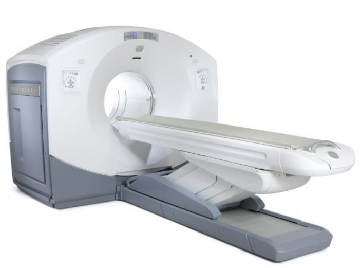
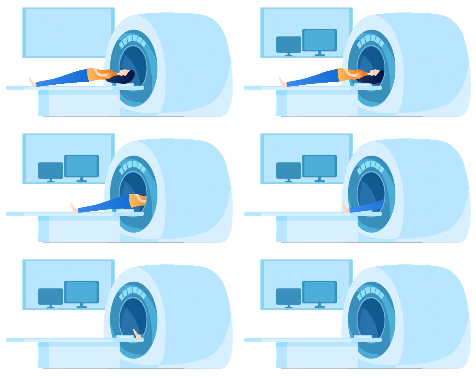
the medical imaging field, the examination that can be called
"top-notch" is none other than PET/CT! This cutting-edge imaging
technology combines PET (precisely capturing metabolic changes in lesions) with
CT (clearly presenting human anatomical structures), forming a powerful imaging
diagnostic capability.

When it comes to PET/CT, the first thing that comes to everyones mind is tumor diagnosis. However, its applications are far more extensive. It can assist doctors in detecting abnormalities earlier, making more accurate disease assessments, and obtaining more comprehensive information in the screening of cardiovascular and cerebrovascular diseases, evaluation of the endocrine system, and differentiation of infections and inflammation.
Our editor conducted an exclusive interview with Professor Yu Lijuan, a renowned imaging diagnosis expert in China and the director of the Department of Nuclear Medicine at Hainan Cancer Hospital, to help everyone systematically understand the various applications of nuclear medicine PET/CT examinations.
Tumor diagnosis and treatment
Four core applications of PET/CT
PET/CT examination plays a pivotal role in different stages of tumor diagnosis and treatment, and is widely recognized as one of the best methods for assisting in tumor diagnosis and treatment decision-making.
1. Preliminary "risk assessment" of suspicious tumors:
When encountering suspicious lesions that are difficult to diagnose, PET/CT can indicate whether there is a malignant tendency by assessing whether the lesion exhibits "high metabolic activity".
2. "Precision profiling" after tumor diagnosis:
After a tumor diagnosis is confirmed, PET/CT can provide a comprehensive understanding of the lesions involvement range through an integrated whole-body scan in one go. Whether its common solid tumors such as lung cancer, prostate cancer, esophageal cancer, and colorectal cancer, or tumors prone to systemic metastasis like lymphoma and melanoma, PET/CT can assist in accurately determining the staging and assessing the status of metastasis.
3. "Efficacy verification" and recurrence monitoring after treatment:
Does the tumor still "survive" after treatment? Are the abnormalities shown in the imaging scars left after treatment or tumor recurrence? PET/CT can clearly determine whether the tissue still has metabolic activity. Compared to examinations that only observe anatomical morphology, it can detect problems earlier, neither missing signals of tumor recurrence nor misidentifying normal scars as lesions.
4. Diagnosis of rare tumors:
Neuroblastoma, neuroendocrine tumors, and other such diseases are often difficult to identify through conventional imaging due to their unique lesion characteristics. However, PET/CT can provide crucial support for the diagnosis of these rare tumors by accurately detecting metabolic abnormalities.

heart disease
"Dont just check whether the blood vessels are blocked or not"
"Further exploring whether the myocardial function is good or not"
Many heart issues are difficult to fully clarify solely through electrocardiogram or coronary CT, especially subtle changes in heart function. However, PET/CT examination can precisely fill this gap:
1. Preoperative "myocardial viability assessment":
Before considering performing a stent or bypass surgery on a patient, myocardial perfusion/metabolic imaging can accurately distinguish the myocardial status: determining which areas are only temporarily ischemic and still have the potential to be "revived", and which have developed into necrotic tissue, providing crucial evidence for whether surgery is necessary and how to proceed.

2. Monitoring of inflammatory heart conditions:
For conditions such as myocarditis and post-heart-transplant rejection, FDG PET/CT can assist in the diagnosis of viral myocarditis, identify myocardial fibrosis, or detect post-heart-transplant rejection promptly by tracking the accumulation of inflammatory cells, thereby facilitating early intervention for such complex cardiac diseases.
nervous system
Abnormal positioning, earlier disease detection
Metabolic abnormalities in the brain often occur earlier than structural changes, and PET/CT, with its keen ability to capture metabolic changes, plays a unique role in the diagnosis and treatment of neurological diseases such as Alzheimers disease:
Many people mistakenly believe that Alzheimers disease begins with memory decline, but in reality, the brains metabolism has already begun to decline subtly before noticeable symptoms appear. PET scans can detect these metabolic abnormalities 15 to 20 years in advance, allowing for valuable time to be gained for ultra-early identification and intervention of the disease.

other aspects
Infection localization, immune monitoring
1. "Precise localization" of infection and inflammation
For issues such as osteomyelitis and artificial joint infections, PET/CT can clearly pinpoint the areas of active inflammation. It is particularly suitable for patients who have undergone repeated treatments without success and whose lesions are concealed, helping to identify the "root cause" and avoid blind medication or treatment.
2. "Monitoring and evaluation" of immune-related diseases
In the diagnosis and treatment of autoimmune diseases such as systemic lupus erythematosus and rheumatoid arthritis, PET/CT also plays an active role. It can assist in assessing the activity level of the disease and determining the distribution range of lesions by detecting areas of active inflammation in the body. Furthermore, by comparing images at different time points, it can dynamically evaluate the therapeutic effect and provide intuitive evidence for adjusting the treatment plan.

As a Class I clinical key specialty in our province, the Department of Nuclear Medicine at Hainan Cancer Hospital not only brings together a team of domestic first-class and provincial-leading experts, but also establishes a rare integrated multi-modal imaging diagnosis and radionuclide therapy center in China, focusing on "imaging + nuclear medicine examination", providing strong technical support for the precise diagnosis and treatment of tumors and various diseases.
Expert Introduction

Yu Lijuan
Director of Nuclear Medicine Department, Medical Imaging Department
Chief Physician, Professor
Postdoctoral researcher, supervisor of graduate students
Doctoral supervisor, top-notch talent in Hainan Province
Provincial Outstanding Science and Technology Worker
Medical expertise
Multimodal imaging diagnosis of tumors includes X-ray, CT, MR, SPECT/CT, and PET/CT, etc. I am particularly skilled in the differential diagnosis of small nodular lung cancer and the PET/CT diagnosis of malignant tumors in various systems throughout the body.
Clinic Hours
Multidisciplinary Clinic for Thoracic Tumors
Friday morning
Pulmonary nodule outpatient clinic
Monday morning, Tuesday morning, Wednesday morning
Nuclear medicine outpatient clinic
Monday to Friday
Written by | Chen Lin

